
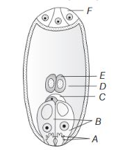
Identify A to F in the diagram given below.
Answer & explanation
Correct answer: option 1
The correct answer is Option (1) → A–Filiform apparatus, B–Synergid, C–Egg, D–Central cell, E–Polar nuclei, F–Antipodals
The mature embryo sac is 7-celled and 8-nucleate. The micropylar end is at the bottom of the diagram and the chalazal end at the top.
Reading the diagram from the bottom: A is the finger-like thickening at the tip of the synergids, the filiform apparatus. B marks the two synergids. C is the egg, placed just above the synergids. D is the large central cell, and E marks the two polar nuclei inside it. F marks the three antipodals at the top. Only Option (1) gives all six labels in this order.
NCERT Extract:
The Pistil, Megasporangium (Ovule) and Embryo Sac – “… Observe the distribution of cells inside the embryo sac (Figure 1.8b, c). Six of the eight nuclei are surrounded by cell walls and organised into cells; the remaining two nuclei, called polar nuclei are situated below the egg apparatus in the large central cell. There is a characteristic distribution of the cells within the embryo sac. Three cells are grouped together at the micropylar end and constitute the egg apparatus. The egg apparatus, in turn, consists of two synergids and one egg cell. The synergids have special cellular thickenings at the micropylar tip called filiform apparatus, which play an important role in guiding the pollen tubes into the synergid. Three cells are at the chalazal end and are called the antipodals.”
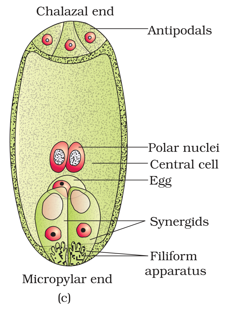
Figure 1.8 (c) A diagrammatic representation of the mature embryo sac