
In Sickle-cell anemia the substitution of amino acid in the globin protein results due to the single base substitution at the sixth codon of the β-globin gene from _____.
Answer & explanation
Correct answer: option 2
The correct answer is Option (2) → GAG to GUG
A codon is a group of three bases on the mRNA that stands for one amino acid. In the normal β-globin gene, the sixth codon is GAG, which codes for glutamic acid.
Option (1) GAG to AUG — Incorrect. The change from GAG to AUG needs two bases to change, not one. AUG codes for methionine and is also the start codon.
Option (2) GAG to GUG — Correct. Only the middle base changes, A to U. The new codon GUG codes for valine, so valine takes the place of glutamic acid in the globin chain.
Option (3) CAG to AUG — Incorrect. The starting codon is wrong. CAG codes for glutamine, not glutamic acid, and the change to AUG would again need two bases.
Option (4) UAG to GUG — Incorrect. UAG is a stop codon. It codes for no amino acid, so it cannot be the sixth codon of the gene.
Note: A similar question was also asked in CUET (UG) 2025 on 20-05-2025 (Slot 1).
NCERT Extract:
Mendelian Disorders – “… Sickle-cell anaemia : This is an autosome linked recessive trait that can be transmitted from parents to the offspring when both the partners are carrier for the gene (or heterozygous). The disease is controlled by a single pair of allele, HbA and HbS. Out of the three possible genotypes only homozygous individuals for HbS (HbSHbS) show the diseased phenotype. Heterozygous (HbAHbS) individuals appear apparently unaffected but they are carrier of the disease as there is 50 per cent probability of transmission of the mutant gene to the progeny, thus exhibiting sickle-cell trait (Figure 4.15). The defect is caused by the substitution of Glutamic acid (Glu) by Valine (Val) at the sixth position of the beta globin chain of the haemoglobin molecule. The substitution of amino acid in the globin protein results due to the single base substitution at the sixth codon of the beta globin gene from GAG to GUG.”
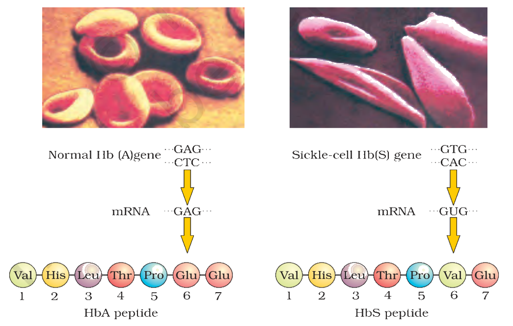
Figure 4.15 Micrograph of the red blood cells and the amino acid composition of the relevant portion of β-chain of haemoglobin: (a) From a normal individual; (b) From an individual with sickle-cell anaemia