
Gel electrophoresis
Read the following passage carefully and answer the given questions.
The cutting of DNA by molecular scissors results in fragments of DNA. These fragments can be separated by a technique known as gel electrophoresis. Since DNA fragments are negatively charged molecules, they can be separated by forcing them to move toward electrode under an electric field through a matrix. Nowadays, most commonly used matrix is agarose, which is a natural polymer. The DNA fragments resolve according to their size through sieving effect provided by agarose gel. Hence, smaller fragment size, farther it moves. The separated DNA fragments can be visualized only after staining DNA with a dye followed by exposure to radiation. The separated bands of DNA are cut out from agarose gel and extracted from gel piece. The DNA fragments purified in this way are used in construction recombinant DNA by joining them with cloning vectors.
In gel electrophoresis, DNA can be visualized as orange bands in the presence of:
Answer & explanation
Correct answer: option 3
The correct answer is Option (3) → Ultraviolet light
Pure DNA fragments cannot be seen in visible light. The gel is stained with ethidium bromide. This dye binds to the DNA. When the gel is then exposed to UV radiation, the bound dye glows, and the DNA shows up as bright orange bands.
(1) Sun light — Incorrect. This is visible light, in which the stained bands do not show up.
(2) Fluorescent light — Incorrect. A fluorescent lamp gives only visible light. It lacks the UV needed to make the dye glow.
(3) Ultraviolet light — Correct. Ultraviolet light is the UV radiation of the book. It makes the ethidium bromide glow orange.
(4) Infrared light — Incorrect. Infrared does not make the dye glow.
Note: The passage is taken from NCERT. NTA has put lines of the NCERT text together to make it. Most CUET (UG) questions come straight from NCERT, so study the book line by line, including figures and their titles.
NCERT Extract:
Restriction Enzymes – …
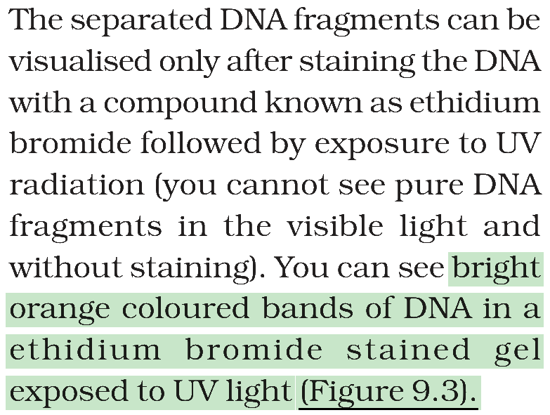
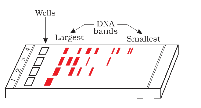
Figure 9.3 A typical agarose gel electrophoresis showing migration of undigested (lane 1) and digested set of DNA fragments (lane 2 to 4)