
Which of the following options correctly represents the sequence of parts in an ovule, starting from the innermost part?
Answer & explanation
Correct answer: option 4
The correct answer is Option (4) → Egg, embryo sac, nucellus, integument
An ovule is built in layers. The integuments form the outer protective envelopes. Inside them is the nucellus, a mass of food-rich cells. The embryo sac (female gametophyte) lies within the nucellus, and the egg cell is one of the cells of the egg apparatus inside the embryo sac. From the inside out, the order is egg, embryo sac, nucellus, integument.
Option (1) Embryo sac, nucellus, integument, egg — Incorrect. The egg lies inside the embryo sac, so it cannot be the outermost part. Starting with the embryo sac also skips the egg.
Option (2) Egg, nucellus, embryo sac, integument — Incorrect. The embryo sac lies inside the nucellus, not outside it, so the nucellus cannot come before the embryo sac.
Option (3) Egg, integument, embryo sac, nucellus — Incorrect. The integuments are the outermost envelopes of the ovule. They enclose the nucellus and cannot sit between the egg and the embryo sac.
Option (4) Egg, embryo sac, nucellus, integument — Correct. Egg (in the egg apparatus) → embryo sac → nucellus → integument is the correct order from the innermost part outwards.
NCERT Extract:
The Pistil, Megasporangium (Ovule) and Embryo Sac – “… Each ovule has one or two protective envelopes called integuments. Integuments encircle the nucellus except at the tip where a small opening called the micropyle is organised. Opposite the micropylar end, is the chalaza, representing the basal part of the ovule. Enclosed within the integuments is a mass of cells called the nucellus. Cells of the nucellus have abundant reserve food materials. Located in the nucellus is the embryo sac or female gametophyte.”
The Pistil, Megasporangium (Ovule) and Embryo Sac – “… Three cells are grouped together at the micropylar end and constitute the egg apparatus. The egg apparatus, in turn, consists of two synergids and one egg cell.”
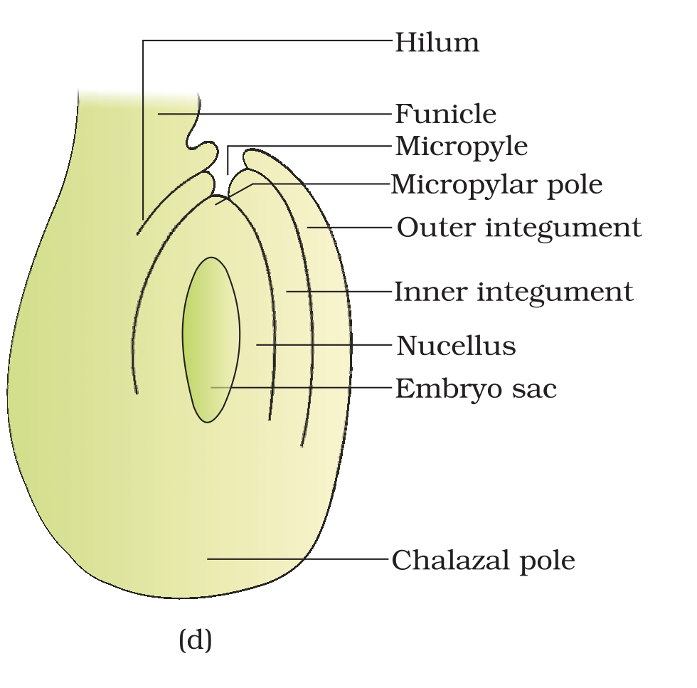
Figure 1.7 (d) A diagrammatic view of a typical anatropous ovule