
What is the filiform apparatus in the embryo sac of angiosperms?
Answer & explanation
Correct answer: option 2
The correct answer is Option (2) → Special cellular thickenings of the synergids at the micropylar tip
The mature embryo sac is 7-celled and 8-nucleate. At its micropylar end lies the egg apparatus, made of two synergids and one egg cell. Each synergid carries finger-like thickenings at its micropylar tip, and these together form the filiform apparatus. It guides the pollen tube into a synergid.
Option (1) Special cellular thickenings at antipodal cells — Incorrect. The antipodals are the three cells at the chalazal end of the embryo sac, far from the micropyle. They carry no such thickenings.
Option (2) Special cellular thickenings of the synergids at the micropylar tip — Correct. The filiform apparatus is the set of special cellular thickenings of the synergids at the micropylar tip. It plays an important role in guiding the pollen tube into the synergid.
Option (3) Special cellular thickenings of endosperm — Incorrect. The endosperm is a food-storing tissue that forms after fertilisation, from the primary endosperm nucleus. It is not a part of the mature embryo sac.
Option (4) Special cellular thickenings of egg cell — Incorrect. The egg cell lies between the two synergids at the micropylar end, but the thickenings belong to the synergids, not to the egg.
NCERT Extract:
The Pistil, Megasporangium (Ovule) and Embryo Sac – “… There is a characteristic distribution of the cells within the embryo sac. Three cells are grouped together at the micropylar end and constitute the egg apparatus. The egg apparatus, in turn, consists of two synergids and one egg cell. The synergids have special cellular thickenings at the micropylar tip called filiform apparatus, which play an important role in guiding the pollen tubes into the synergid. Three cells are at the chalazal end and are called the antipodals.”
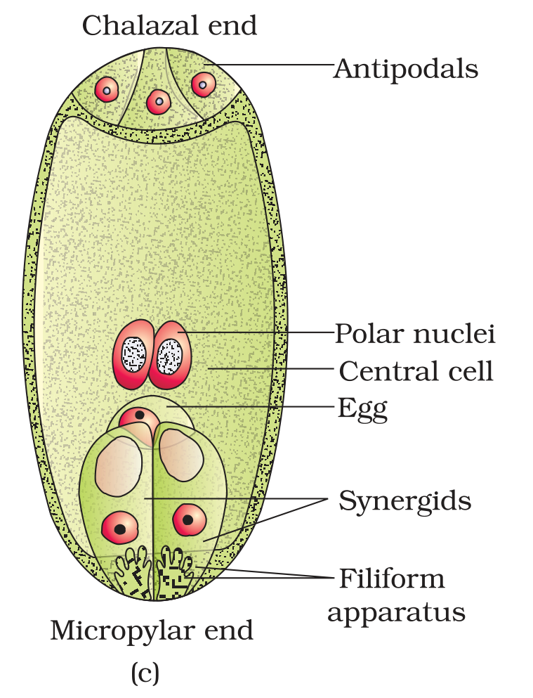
Figure 1.8 (c) A diagrammatic representation of the mature embryo sac