
Arrange the given steps involved in gel electrophoresis used for separation of DNA fragments?
(A) Exposure to UV light
(B) Staining with ethidium bromide
(C) Movement of DNA fragments towards anode
(D) Elution
Choose the correct answer from the options given below.
Answer & explanation
Correct answer: option 4
The correct answer is Option (4) → (C), (B), (A), (D)
(C) Movement of DNA fragments towards anode — first. DNA is negatively charged. In the electric field the fragments move through the agarose gel towards the anode, and the smaller ones move farther.
(B) Staining with ethidium bromide — second. Pure DNA cannot be seen in visible light. So the gel is stained after the run.
(A) Exposure to UV light — third. Under UV light the stained DNA shows up as bright orange bands.
(D) Elution — fourth. The band of the wanted fragment is cut out and the DNA is extracted from the gel piece. This last step is elution.
Note: The same question was also asked in CUET (UG) 2025 on 13-05-2025 (Slot 1).
NCERT Extract:
Restriction Enzymes – “… Separation and isolation of DNA fragments : The cutting of DNA by restriction endonucleases results in the fragments of DNA. These fragments can be separated by a technique known as gel electrophoresis. Since DNA fragments are negatively charged molecules they can be separated by forcing them to move towards the anode under an electric field through a medium/matrix. Nowadays the most commonly used matrix is agarose which is a natural polymer extracted from sea weeds. The DNA fragments separate (resolve) according to their size through sieving effect provided by the agarose gel. Hence, the smaller the fragment size, the farther it moves. Look at the Figure 9.3 and guess at which end of the gel the sample was loaded. The separated DNA fragments can be visualised only after staining the DNA with a compound known as ethidium bromide followed by exposure to UV radiation (you cannot see pure DNA fragments in the visible light and without staining). You can see bright orange coloured bands of DNA in a ethidium bromide stained gel exposed to UV light (Figure 9.3). The separated bands of DNA are cut out from the agarose gel and extracted from the gel piece. This step is known as elution.”
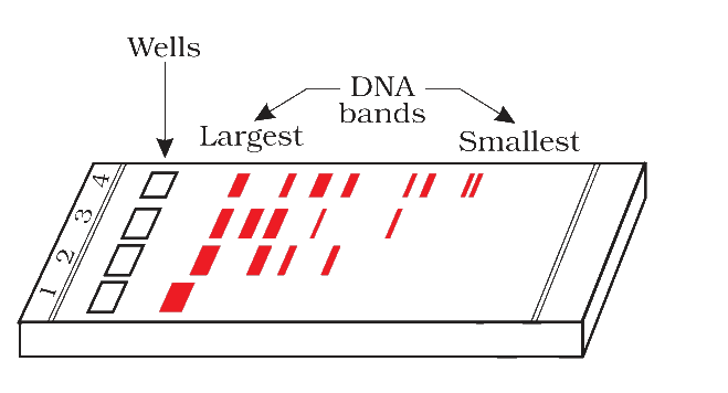
Figure 9.3 A typical agarose gel electrophoresis showing migration of undigested (lane 1) and digested set of DNA fragments (lane 2 to 4)