
Which disorder is caused by the substitution of Glutamic acid (Glu) by Valine (Val) at the sixth position of the beta globin chain of the haemoglobin ?
Answer & explanation
Correct answer: option 2
The correct answer is Option (2) → Sickle-cell Anaemia
Option (1) Phenylketonuria — Incorrect. It is an inborn error of metabolism. The person lacks the enzyme that changes phenylalanine into tyrosine, and phenylalanine collects in the brain.
Option (2) Sickle-cell Anaemia — Correct. A single base change in the gene, GAG to GUG, puts valine in place of glutamic acid. The faulty haemoglobin makes the RBCs sickle-shaped at low oxygen.
Option (3) Haemophilia — Incorrect. It is a sex-linked recessive disease. A protein of the chain of blood clotting is affected, so even a small cut keeps bleeding.
Option (4) Thalassemia — Incorrect. Thalassemia is also a disorder of haemoglobin, but it is a quantitative problem: too few globin chains are made. Sickle-cell anaemia is a qualitative problem: the globin is made, but with a wrong amino acid.
Note: A similar question was also asked in CUET (UG) 2023 on 28-05-2023 (Slot 1).
NCERT Extract:
Mendelian Disorders – “… Sickle-cell anaemia : This is an autosome linked recessive trait that can be transmitted from parents to the offspring when both the partners are carrier for the gene (or heterozygous). The disease is controlled by a single pair of allele, HbA and HbS. Out of the three possible genotypes only homozygous individuals for HbS (HbSHbS) show the diseased phenotype. Heterozygous (HbAHbS) individuals appear apparently unaffected but they are carrier of the disease as there is 50 per cent probability of transmission of the mutant gene to the progeny, thus exhibiting sickle-cell trait (Figure 4.15). The defect is caused by the substitution of Glutamic acid (Glu) by Valine (Val) at the sixth position of the beta globin chain of the haemoglobin molecule. The substitution of amino acid in the globin protein results due to the single base substitution at the sixth codon of the beta globin gene from GAG to GUG.”
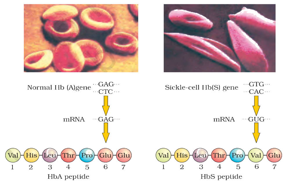
Figure 4.15 Micrograph of the red blood cells and the amino acid composition of the relevant portion of β-chain of haemoglobin: (a) From a normal individual; (b) From an individual with sickle-cell anaemia