
In gel electrophoresis, the separated DNA fragment can be visualized -
Answer & explanation
Correct answer: option 1
The correct answer is Option (1) → When ethidium bromide exposed to UV light
Option (1) When ethidium bromide exposed to UV light — Correct. Pure DNA cannot be seen in visible light. The gel is stained with ethidium bromide and then exposed to UV (ultraviolet) light, and the DNA shows up as bright orange bands.
Option (2) When ethidium bromide exposed to infrared light — Incorrect. The stain is right, but the light is wrong. Ethidium bromide glows only under UV light; infrared light shows nothing.
Option (3) When bromophenol blue exposed to UV light — Incorrect. UV means ultraviolet. Bromophenol blue is a dye added to the sample to follow how far the run has gone. It does not bind DNA, so it cannot show the DNA bands.
Option (4) When acetocarmine exposed to UV light — Incorrect. UV means ultraviolet. Acetocarmine is a stain used to see chromosomes under the light microscope, for example in an onion root tip. It is not used for DNA in a gel.
NCERT Extract:
Restriction Enzymes – “… The separated DNA fragments can be visualised only after staining the DNA with a compound known as ethidium bromide followed by exposure to UV radiation (you cannot see pure DNA fragments in the visible light and without staining). You can see bright orange coloured bands of DNA in a ethidium bromide stained gel exposed to UV light (Figure 9.3).”
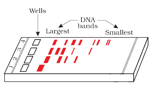
Figure 9.3 A typical agarose gel electrophoresis showing migration of undigested (lane 1) and digested set of DNA fragments (lane 2 to 4)