
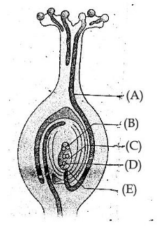
The figure given above shows the growth of pollen tube. Observe the figure and answer the following question.
Identify the part (A) and (B).
Answer & explanation
Correct answer: option 3
The correct answer is Option (3) → Pollen tube and antipodal cells respectively
In the figure, A is the tube growing down from the stigma through the style into the ovule: the pollen tube. B is at the chalazal end of the embryo sac, opposite the micropyle where the tube enters: the antipodal cells.
Option (1) Pollen tube and synergids respectively — Incorrect. B is at the chalazal end; synergids lie at the micropylar end.
Option (2) Synergids and female gamete respectively — Incorrect. The synergids lie with the egg at the micropylar end; they are not the long tube A that grows down the style. A is the pollen tube, and B, at the chalazal end, is the antipodal cells.
Option (3) Pollen tube and antipodal cells respectively — Correct. In the figure, (A) is the pollen tube growing into the embryo sac through the micropyle. (B) shows the three cells at the chalazal end of the embryo sac, the antipodal cells.
Option (4) Pollen tube and female gamete respectively — Incorrect. The female gamete lies at the micropylar end, not at B.