
The centre of each microsporangium is occupied by:
Answer & explanation
Correct answer: option 1
The correct answer is Option (1) → Sporogenous tissue
A young anther in cross-section shows four microsporangia. Each microsporangium has four wall layers on the outside: epidermis, endothecium, middle layers and, innermost, the tapetum. The space in the middle is filled by a group of compactly arranged, similar cells called the sporogenous tissue.
Option (1) Sporogenous tissue — Correct. The sporogenous tissue occupies the centre of each microsporangium. Its cells later divide by meiosis to form microspore tetrads, which develop into pollen grains.
Option (2) Tapetum — Incorrect. The tapetum is the innermost wall layer that surrounds the sporogenous tissue. It lines the centre but does not fill it, and its job is to nourish the developing pollen grains.
Option (3) Nucellus — Incorrect. The nucellus is a mass of cells inside the ovule, the female megasporangium. It holds the embryo sac and is not part of the anther.
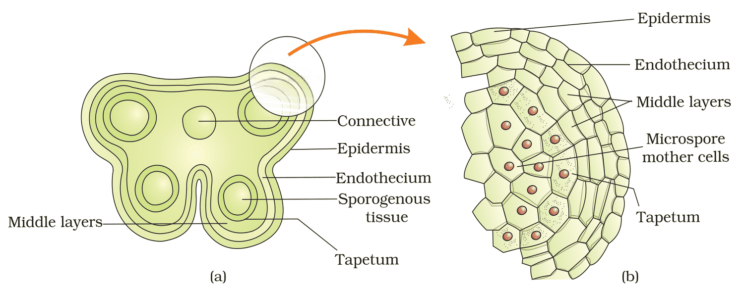
Figure 1.3 (a) Transverse section of a young anther; (b) Enlarged view of one microsporangium showing wall layers
NCERT Extract:
Stamen, Microsporangium and Pollen Grain – “… The innermost wall layer is the tapetum. It nourishes the developing pollen grains. Cells of the tapetum possess dense cytoplasm and generally have more than one nucleus. … When the anther is young, a group of compactly arranged homogenous cells called the sporogenous tissue occupies the centre of each microsporangium.”