
The figure is a diagrammatic representation of protein structure, in which each coloured ball represents an amino acid.
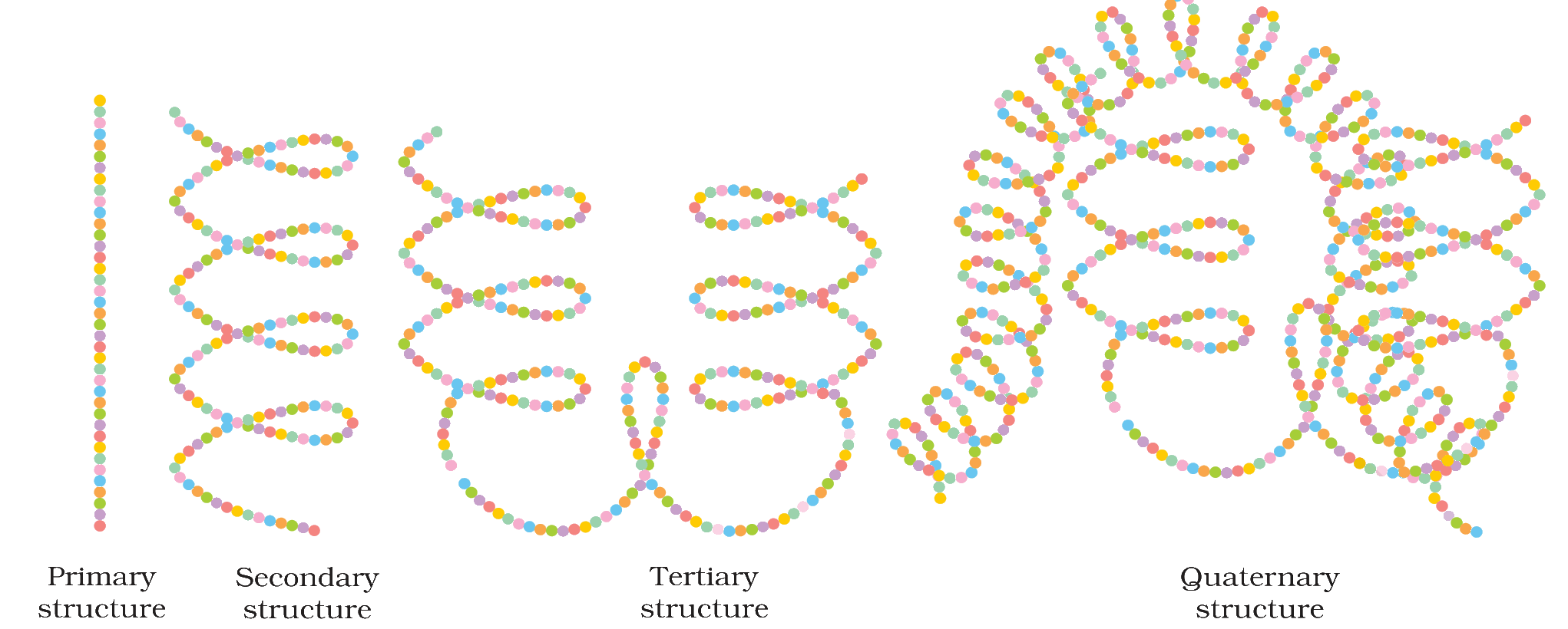
Which type of secondary structure is shown in the figure, and what holds this structure together?
Answer & explanation
Correct answer: option 4
The correct answer is Option (4) → α-Helix; hydrogen bonds between the –NH and >C=O groups of adjacent turns
The secondary structure of a protein exists in two types, the α-helix and the β-pleated sheet. In the figure, the chain labelled “Secondary structure” is a single chain twisted into a regular coil (a screw), so it is an α-helix.
In an α-helix, the –NH group of each amino acid residue is hydrogen bonded to the >C=O group of an adjacent turn of the helix.
NCERT (2026-27 edition):
10.2.3 Structure of Proteins – “(ii) Secondary structure of proteins: The secondary structure of protein refers to the shape in which a long polypeptide chain can exist. They are found to exist in two different types of structures viz. α-helix and β-pleated sheet structure. These structures arise due to the regular folding of the backbone of the polypeptide chain due to hydrogen bonding between >C=O and –NH– groups of the peptide bond.”
“α-Helix is one of the most common ways in which a polypeptide chain forms all possible hydrogen bonds by twisting into a right handed screw (helix) with the –NH group of each amino acid residue hydrogen bonded to the >C=O of an adjacent turn of the helix as shown in Fig.10.1.”
“In β-pleated sheet structure all peptide chains are stretched out to nearly maximum extension and then laid side by side which are held together by intermolecular hydrogen bonds.”
Fig. 10.3: Diagrammatic representation of protein structure (two sub-units of two types in quaternary structure)