What are seen as "beads on string" structure when viewed under an electron microscope?
Answer & explanation
Correct answer: option 4
The correct answer is Option (4) → Nucleosomes
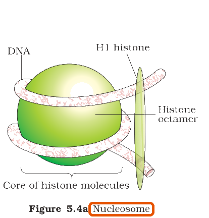
In eukaryotes, the negatively charged DNA is wrapped around a positively charged histone octamer. Each such unit, with about 200 bp of DNA, is a nucleosome. Under the electron microscope a chromatin fibre shows the nucleosomes like beads on a string.
Option (1) Nucleoid — Incorrect. The nucleoid is the region in a prokaryotic cell where its DNA lies, held in loops by proteins.
Option (2) Chromatin — Incorrect. Chromatin is the string itself, the whole fibre; the beads are the nucleosomes on it.
Option (3) Base pairs — Incorrect. Base pairs are far too small to be seen as beads.
Option (4) Nucleosomes — Correct. DNA wrapped around a histone octamer forms a nucleosome. Under the electron microscope chromatin shows these nucleosomes as repeating units, seen as the ‘beads-on-string’ structure.
NCERT Extract:
Packaging of DNA Helix – “… The negatively charged DNA is wrapped around the positively charged histone octamer to form a structure called nucleosome (Figure 5.4 a). A typical nucleosome contains 200 bp of DNA helix. Nucleosomes constitute the repeating unit of a structure in nucleus called chromatin, thread-like stained (coloured) bodies seen in nucleus. The nucleosomes in chromatin are seen as ‘beads-on-string’ structure when viewed under electron microscope (EM) (Figure 5.4 b).”